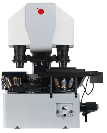
Q-Phase

Q-Phase

The Q-Phase microscope is an advanced holographic microscope for Quantitative Phase Imaging (QPI), designed for label-free, high-sensitivity live-cell imaging. By combining coherence-controlled holography with LED illumination, Q-Phase eliminates common laser-based artifacts, enabling precise quantification of cell dry mass, morphology, and dynamics in real time.

For More Info: Download Datasheet

Core Technology

  • Based on Quantitative Phase Imaging (QPI) with patented coherence-controlled holographic microscopy.

  • Uses an incoherent LED light source (λ = 660 nm) → eliminates coherence noise and artifacts common with lasers.

  • Calculates cell dry mass (pg/μm²) from detected phase shifts, allowing real-time quantitative assessment of living cells.

Performance & Imaging

  • Label-free live-cell imaging → non-invasive, minimal sample manipulation, extremely low phototoxicity.

  • High sensitivity: phase detection down to 0.011 rad.

  • Supports multi-modal imaging: QPI, simulated DIC, brightfield, high-pass filtered phase, and optional fluorescence.

  • Segmentation & tracking: accurate detection of transparent cell boundaries and automated cell profiling.

  • Imaging in scattering/turbid media (e.g., collagen, Matrigel) – unique capacity in QPI.

  • Acquisition speed: 16 fps (higher optional).

Automation & Analysis

  • Fully motorized stage with stage-top incubator for long-term live-cell experiments (temperature, CO₂, humidity control).

  • Advanced SophiQ software: real-time segmentation, data visualization, multi-parameter analysis, gating, and subpopulation tracking.

  • Parameters quantified: cell dry mass, density, area, growth rate, motility (speed, Euclidean distance, meandering index), confluence, fluorescence.

Specifications

  • Objectives: 4× to 60× magnification.

  • Resolution: 0.58 μm (60× NA 1.4 objective).

  • Field of view: up to 1.48 mm × 1.48 mm (4× objective).

  • Stage piezo travel: 500 μm.

  • Microscope footprint includes anti-vibration platform, control panel, and optional fluorescence module with LED illuminator & filter cubes.

Applications

  • Cell biology & cancer research: morphology, drug toxicity, cell cycle, rare event detection.

  • Stem cell research: non-invasive monitoring of differentiation.

  • Immunology & virology: label-free infection and immune response analysis.

  • Pharma & drug screening: cytotoxicity assays, cell viability, nanocarrier uptake.

  • Biomaterials: refractive index mapping of condensates and tissues (e.g., Limulus cornea).

Global Use

  • Installed at leading institutes:

    • Max Planck Institute of Molecular Cell Biology & Genetics (Germany)

    • Masaryk University (Czech Republic)

    • Institute of Molecular Genetics ASCR (Czech Republic)

    • Brno University of Technology (Czech Republic)