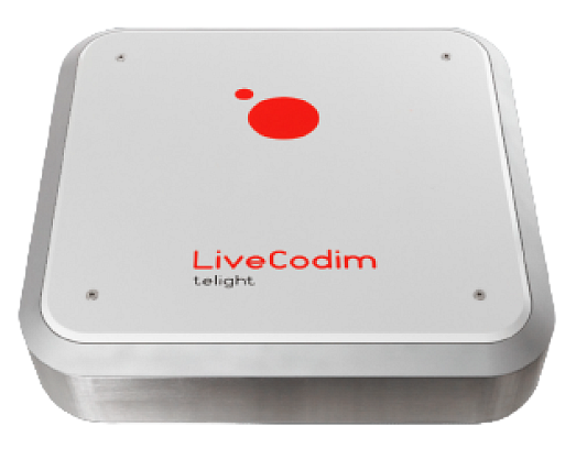
LiveCodim

LiveCodim

LiveCodim transforms an existing inverted microscope into a flexible widefield–confocal–super-resolution system that delivers ~90 nm lateral resolution while minimizing phototoxicity and photobleaching, making it well-suited for live-cell imaging. Using conical diffraction from a biaxial crystal to generate structured illumination patterns, it reconstructs super-resolved images across up to four laser channels, with confocal-like depth reach (to ~500 μm). The GPU-accelerated software streamlines acquisition and processing, outputs OME-TIFF, and integrates smoothly with ImageJ/Imaris. Because it’s achromatic, modular, and microscope-agnostic, LiveCodim is a practical way to add high-performance super-resolution to standard confocal workflows—without changing sample prep.

For More Info: Download Datasheet

What it is

  • 3-in-1 imaging platform that upgrades a standard widefield microscope to widefield, confocal, and super-resolution modes.

  • Based on conical diffraction with a biaxial crystal to create structured illumination; scans patterns in three orientations for super-resolved reconstructions.

Key capabilities

  • Gentle super-resolution: up to ~90 nm lateral (at 488 nm) with low phototoxicity and little-to-no photobleaching.

  • Depth penetration comparable to confocal; imaging up to ~500 μm (with long-WD objectives).

  • Multicolor: up to 4 laser lines (405/488/561/640 nm; customizable).

  • Switch modes with one click; no change in sample prep vs. confocal.

Compatibility & integration

  • Achromatic & modular (400–1000 nm); fits any major inverted microscope with left/right side port (e.g., Nikon Ti, Zeiss AxioObserver, Evident IX83, Leica DMi…).

  • Works with incubators/CO₂ fittings, standard LED widefield illumination, filter turrets/cubes, high-NA oil objectives (60×/63×, NA 1.40–1.49), motorized Z and XY.

  • Outputs OME-TIFF; TIFF for images.

Software & workflow

  • Intuitive UI for acquire/process/visualize; GPU-accelerated reconstruction; adaptive SR processing.

  • FAIR data; direct export to ImageJ & Imaris; communicates with open-source tools.

Performance & specs

  • Lateral resolution: up to ~90 nm (488 nm, high-NA oil).

  • Axial resolution: up to ~500 nm (488 nm, high-NA oil).

  • Max scanned FOV: 60 × 60 μm (60× objective).

  • Acquisition speed: ~2 s per 10 × 10 μm area.

  • Camera: sCMOS (≈96% QE).

  • Operation: point scanning with structured PSF; variable pinhole.

  • Footprint: ~52 × 48 × 14.5 cm, ~20 kg.

Applications

  • Cell biology, cytoskeleton dynamics, cell cycle, cancer biology, microbiology, neurobiology, plant biology—and broadly “your sample”.

Why it’s different

  • True optical super-resolution (not just deconvolution) with low-intensity illumination window → preserves live cells.

  • Confocal-like geometry + structured illumination → SR detail with deeper scanning.

No special dyes/probes beyond standard confocal labeling; no exotic sample prep.